Workshop “Collective motion of active swimmers”
September 25-26, 2013
Université de Nice Sophia Antipolis, Nice
Purpose of the workshop
The goal of the workshop is to provide an interdisciplinary forum gathering biologists, mathematicians and physicist working on the experimental observation and numerical modeling of suspensions of active swimmers. The workshop will consist of 6 one-hour lectures by experts and 8 thirty-minutes more specialized presentations.
Meeting dates and location
The meeting dates are:
- Wednesday 25 September 2013,
- Thursday 26 September 2013.
The location of the meeting is at
Laboratoire Jean-Alexandre Dieudonné
Université de Nice Sophia Antipolis
Parc Valrose, 28 Avenue Valrose
06108 Nice Cedex 02
Maps :
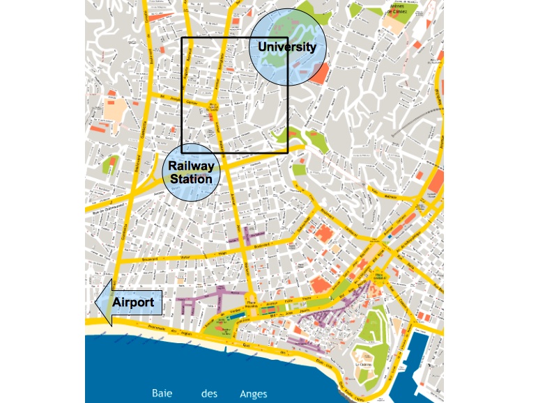
- Map of Nice
- Interactive campus map
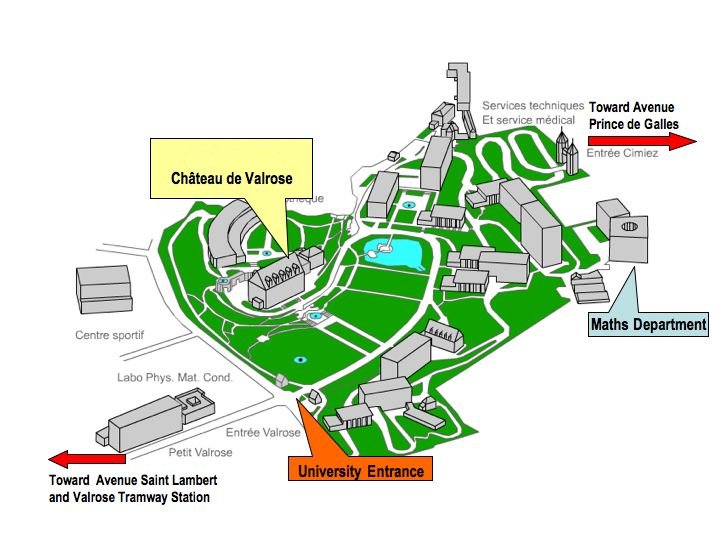
- campus map (Lab. Dieudonné = maths department)
For directions, see below
Sponsors
The workshop is sponsored by the ANR project ‘MOTIMO’.
MOTIMO is an acronym for ‘Seminal Motility Imaging and Modeling’.
Scientific committee
- Laure Blanc-Féraud, Laboratoire d’Informatique Signaux et Systèmes (I3S) of Sophia-Antipolis
- Pierre Degond, Institut de Mathématiques de Toulouse
- Xavier Druart, Laboratoire de Physiologie de la Reproduction et du Comportement of INRA-Tours
- Charles Kervrann, INRIA Rennes
- Enkeleida Lushi, Imperial College London
- Philippe Peyla, University Joseph Fourier, Grenoble
- Franck Plouraboué, Institut de Mécanique des Fluides de Toulouse
Local organizing committee and conference web site
Didier Auroux
Laboratoire d’Informatique Signaux et Systèmes (I3S) of Sophia-Antipolis
Conference secretariat
Angelique Guitard
Schedule

Confirmed speakers
One-hour lectures:
Title: Bioconvection and the dispersion of microorganisms in laminar and turbulent flows
Abstract: Many microorganisms swim in preferred directions. For example, many algae swim towards regions of weak light intensity and away from potentially damaging bright-light conditions (termed phototaxis). Even in the dark cells tend to swim upwards (gravitaxis) due to bottom-heaviness or sedimentary torques, a strategy that may be advantageous in deep or murky ponds. This behaviour is modified significantly in shear flows, leading cells to swim towards regions of downwelling fluid, a response called gyrotaxis. Hence, as the microorganisms are typically more dense than the fluid in which they swim, accumulations of cells can drive hydrodynamic instabilities, resulting in ‘bioconvection patterns’ in just tens of seconds over length scales of centimetres. Furthermore, biased swimming across streamlines can lead to drift and diffusion in laminar and turbulent flows that is qualitatively distinct from that of tracers.
Here, I shall describe measurements and theory for individual swimming microorganisms and how one can scale-up to model a suspension of cells and thus bioconvection. I shall discuss the use of differential dynamic microscopy as a high-throughput method to measure attributes of the cells’ swimming motion. And finally, a continuum description will be employed to extend classical Taylor-Aris theory to describe the axial transport of biased swimming cells in flows in tubes and channels. In particular, the results will be used to calculate the dispersion of gyrotactic swimming algae in vertical tubes and channels, results of potential relevance to the algal biofuels industry.
Ecole Normale Supérieure de Lyon
Title: Kinetic theory and hydrodynamic equations for systems of self-propelled particles
Abstract: Hydrodynamic equations governing the density and velocity fields have been derived from the microscopic dynamics, for a gas of self-propelled particles with velocity-aligning binary interactions. The homogeneous state with zero hydrodynamic velocity is unstable above a critical density, signaling the onset of collective motion. While the homogeneous flow is found to be stable far from the transition line, it becomes unstable with respect to finite-wavelength perturbations close to the transition, implying a non-trivial spatio-temporal structure for the resulting flow. Solitary wave solutions of the hydrodynamic equations are found by numerical methods. Considering instead binary interactions with nematic symmetries leads to nematic (rather than polar) order, but the global picture of an unstable homogeneous ordered state close to the onset of order, resulting in non-linear patterns, remains the same. This global picture is further confirmed by eventually considering diffusive active nematic particles.
Title: Surface interactions in suspensions of swimming cells
Abstract: Interactions between swimming cells and surfaces are essential to many microbiological processes, from bacterial biofilm formation to human fertilization. However, despite their fundamental importance, relatively little is known about the physical mechanisms that govern the scattering of flagellated or ciliated cells from solid surfaces. In the talk I will reveal recent advances in understanding of flagella interaction with surfaces, provide mechanisms for utilizing our knowledge about these interactions to control swimming of flagellated cells. In addition, I will describe our very recent results on sperm rheotaxis near surfaces. The key focus will be on the experimental results, supported by numerical simulation using minimal models.
Title: A detection-based framework for sub-cellular trafficking and exocytosis analysis in tirf microscopy
Abstract:
Total Internal Reflection Fluorescence (TIRF) microscopy allows for the imaging of events occuring at a narrow depth (100nm-300nm) from a glass cover slip. Such ability is particularly useful in cell biology when events occuring at or near the membrane, involving communication between the cell and the external medium, are of interest. Those studies typically involve an important number of localised individual events which may be correlated across two channels. For this purpose an EM-CCD camera equiped with a image-splitter was installed on a TIRF microscope, allowing for in vivo simultaneous dual color TIRF acquisition. In this work is presented a framework for such study based on the automated detection of events at the membrane and their analysis.
The biological model under study involves trans-membrane protein endocytosis/recycling pathways. During their recycling, individual vesicles fuse with the plasma membrane. Due to the exponentially decreasing nature of the sensitivity of TIRF microscopy in z, it corresponds to a sudden appearance in the image. The proposed framework is based on a novel statistical patch-based change detection algorithm that allow for the exhaustive detection of appearances. When repeated across several acquisitions, there are enough individual events for a statistical or biophysical analysis of the resulting collection to be relevant.
Using that framework, we compare the recycling of langerin and Transferin receptor (TfR) and show that at least two biophysical mechanisms are responsible for the behaviour of langerin vesicles at the membrane, where TfR ones shows only diffusion. Dual color experiments involving langerin and Rab11, a protein known to be involved in vesicle recycling at the membrane, are also presented, opening the door for other pairwise experiments and allowing for the in vivo study of the spatio-temporal mechanisms of recycling at the membrane.
Laboratoire Interdisciplinaire de Physique, Université Joseph Fourier, Grenoble
Title: The Physics of Plankton
Abstract: Marine and in-land plankton produces half of the oxygen on earth. It is a very important compound of our ecosystem and it can be also cultivated for several applications. Plankton species that are grown in alga-culture are mainly microalgae which are microswimmers. Commercial and industrial algae cultivation has a lot of potential applications, including food, food dyes, omega3 acids, bioplastics, fertilizers, pollution control and algal fuel. The biggest challenges concerning all this applications are the separation, filtration and concentration processes. To this end, we propose to study and develop a new method of separation/concentration based on the control of the hydrodynamics of an algal suspension coupled with phototaxis (when an organism moves in response to the stimulus of light) [1]. Experiments as well as simulations will be presented.
References:
- [1] Xabel Garcia, Salima Rafai and Philippe Peyla “Light control of the flow of phototactic microswimmer suspensions.” Phys. Rev. Lett. (2013) vol. 110, 138106
École Polytechnique Fédérale de Lausanne
Title: Bioimage informatics algorithms: detection, localization, and tracking fluorescent particles
Abstract: The emergence of the time-lapse fluorescence microscopy have allowed the observation and the quantification of molecular dynamics within living cells. The quantification is significant after the analysis of large number of cells in high-throughput screening environment that requires advanced image-analysis tools. These tools mainly involve the detection, the localization and the tracking of fluorescent proteins that appear as small bright spot in images.
In this context, the localization microscopy (PALM/STORM) achieves super-resolution by rendering positions of accurate localizations of single-molecule emitters. For this task, many software have been developed. Here, we summarized a study to benchmark these localization software. To conduct the evaluation, we designed bio-inspired datasets generated with a faithful image formation model and we established fair metrics for the assessment.
In molecular biology, numerous relevant questions are related to the motility and the diffusion of specific proteins. These information are extracted from fluorescence images by tracking algorithms. The tracking task is complicated by the crossing trajectories of similar particles. We adopted a global optimization framework based on the dynamic programming algorithm. This algorithm code the motion model in a merit function. It is able to track single particle in noisy image even when the photons emission is very dim. For certain applications, additional image modality (e.g. phase contrast) is integrated in the image-analysis system, helping to identify the cell.
Finally, we present several implementations of the detection and tracking modules as ImageJ plugins for biological applications: study dynamics of telomeres, recording the circadian gene expression, measure the growth-rate of a colony of bacteria, construction of the cell lineage in FUCCI images, quantification of the asymmetric cell division.
Thirty-minute presentations:
Institut de Mécanique des Fluides de Toulouse
Title: Spectral Analysis of the collective dynamic in ram semen in confined cells
Abstract:
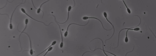
Concentrated swarming flows display turbulent structures at very small Reynolds. In a number of mammal’s species, sperm semen display a collective motion called massal mobility which is associated with whirlpools dynamics observed with phase-contrast microscopy. The understanding of the relaying mecanims of this phenomenon is still an open question. Here we present the first quantitative statistical analysis of the whirlpools spatio-temporal dynamics, collected over large data sets.
We analyze both spatial correlation and velocity distribution of whirlpools, especially from the evaluation of the velocity field obtained by PIV computation of highly concentrated ram semen confined in different cell sizes. We find that the spatial correlation curves collapseon a master curve one the length is rescaled by the integral scale of the velocity field. Furthermore, the spectrum scaled by the energy at the spermatozoon scale wavenumber display an interesting collapse which is the signature of an inverse spectrum cascade.
Laboratoire d’Informatique Signaux et Systèmes (I3S) of Sophia-Antipolis
Title: An introduction to random forests
Abstract: “Machine learning, a branch of artificial intelligence, concerns the construction and study of systems that can learn from data. – Wikipedia.com” An important problem of machine learning is the design of algorithms to perform automatic classification, i.e. the inference without user input of the category an object belongs to among a set of predefined categories. The “object” can be a piece of text, a sound recording, an image, a 3-D mesh… Supervised learning is the process of building an inference rule from a set of samples tagged with their known category, as defined by an expert. The samples themselves are signatures of objects, i.e. synthetic, hopefully discriminative representations of them. Among supervised learning methods, the Random forest procedure has become very popular for its conceptual simplicity and its low computational requirements, and because free, open source implementations are available for several main programming languages. This talk will present the main ideas and properties of Random forests.
Title: Direct numerical simulation of active suspensions
Abstract: It has been observed that, above a certain concentration, the global motion of active suspensions highly differs from the sum of individual motion.
In fact, suspensions of microswimmer have shown to lead to complex dynamics such as the so-called weak turbulence or bioconvection phenomenon.
One of the most spectacular effects pertains to abnormal rheological behavior: the apparent viscosity of an active suspension may be higher or smaller than the
corresponding passive suspension, depending on the type of motility, the cells shape and the concentration.
The link between the individual and the collective behaviour of micro-swimmers is still unperfectly understood.
We aim to develop a numerical tool which is capable of simulating at microscopic level a large quantity of self-propelled particles in a low Reynolds number flow,
retrieving the correct collective motion in the suspension at the macroscopic level. This microscopic approach can be called “direct simulation”.
Swimmers are modelled individually as ellipsoidal rigid particles, and self-propulsion is taken into account through a dipole of forces
distributed on the particle and on the fluid. Hydrodynamic interactions are computed exactly, up to the order of precision of the numerical scheme used to solve the Stokes equations.
We show two dimensional simulations of dilute and dense suspensions of puller and pusher-like micro-swimmers.
Spatial and temporal correlations of the “turbulent” structures appearing when the concentration is high enough are analyzed, as well as the rheological properties of suspensions under shear stress.
Institut de Mécanique des Fluides de Toulouse
Title: Hydrodynamic interactions among large populations of swimming micro-organisms
Abstract: Interplays in many-bodied systems result in intricate patterns, the understanding of which requires an in-depth knowledge of the suspension microstructure and statistics. Representative and reliable statistics require a large number of interacting swimmers (denoted Np) that many simulation methods can hardly afford [e.g. Np ≤ 40 in Mehandia and Nott (2008) or Np = 216 in Ishikawa et al. (2008)]. As a preliminary study, we consider a spherical swimmer model from the classical low Reynolds number framework and implemented in the force-coupling method (FCM) [Climent, Maxey (2009)] for large populations. Resulting statistics reveal non-trivial spatial arrangements of swimmers depending on their swimming gait.
In a second part, if time permits, we will discuss more detailled individual spermatozoon description. More specifically, the precise modelling of the flagellum mechanic properties. Comparisons with the litterature will lead us to a model fitting the computationally efficient FCM strategy for highly populated suspensions.
References:
- Mehandia V, Nott P. 2008. The collective dynamics of selfpropelled particles. J. Fluid Mech. 595 :239–264.
- Ishikawa T, Locsei JT, Pedley TJ. 2008. Development of coherent structures in concentrated suspensions of microswimmers. J Fluid Mech. 615 :401–431.
- The Force Coupling Method : A flexible approach for the simulation of particulate flows, E. Climent & M.R. Maxey, (2009) inserted in “Theoretical Methods for Micro Scale Viscous Flows”, Ressign Press, Eds F. Feuillebois and A. Sellier (ISBN : 978-81-7895-400-4).
Title: Rheological effects on microswimmers in confined geometries
Abstract: Two key factors impacting sperm motility and migration are the rheological properties of the surrounding fluid and the presence of boundaries. In this talk, we will present simulations that explore the relationship between sperm, fluid rheology and boundary features.
Physiological mucus is a suspension of protein filaments that twist, align and recoil with flow generated by beating flagella. These filaments endow mucus with complex properties that affect sperms’ ability to penetrate. Using the method of femlets, a new finite element technique entailing an immersed force representation of the swimmer with a body-fitted mesh, we reveal novel physical mechanisms through which shear-thinning, an important property of physiological mucus affects microscopic swimmers. In particular we show that depending on the stroke employed, microswimmers move either more slowly or more quickly than they would in water. We then show preliminary results for arrays of simple swimmers in confined two-dimensional channels.
Human sperm must also navigate the labyrinthine structure of human fallopian tubes. Sperm trajectories are greatly affected by boundaries, scattering over features such as steps and ripples. We present preliminary work on a mathematical framework, derived from that of Tornberg and Shelley (J. Comp. Phys 196, pp 8-40, 2004) and Gadêlha et al (J. Roy Soc. Int. 7, pp. 1689-97, 2010), for simulating the three-dimensional motion of sperm driven by internally-generated shear forces. The numerical method is fully implicit, geometrically nonlinear and takes into account non-local hydrodynamic effects of boundaries, the cell head and ambient flow. The choice of constant velocity travelling waves of active sliding moment density is informed by flagellar waveforms captured from high-speed digital imaging of live cells penetrating high-viscosity fluid.
Institut de Mathématiques de Toulouse
Title: Hydrodynamic limit and numerical solutions in a system of self-propelled particles
Abstract: We consider an Individual based model of self-propelled particles with alignment interaction and repulsion and study the hydrodynamic limit of this system and its numerical solution. In this model, the particles trend to move alignment with the mean direction of their neighborhoods and try to maintain a minimum distance between themselves. The alignment interaction is supposed to be weakly non-local. This leads to derive a diffusive term in the macroscopic system. Finally, we provide some numerical solutions to the macroscopic system.
Title: Fluorescence microscopy based on compressed sensing
Abstract: In fluorescence microscopy, one can distinguish two kinds of imaging approaches, wide- field and raster scan microscopy, differing by their excitation and detection scheme. In both imaging modalities the acquisition is independent of the information content of the image. Rather, the number of acquisitions N, is imposed by the Nyquist-Shannon theorem. However, in practice, many biological images are compressible (or, equivalently here, sparse), meaning that they depend on a number of degrees of freedom K that is smaller that their size N. Recently, the mathematical theory of compressed sensing (CS) has shown how the sensing modality could take advantage of the image sparsity to reconstruct images with no loss of information while largely reducing the number M of acquisition.
Here we present a novel fluorescence microscope designed along the principles of CS. It uses a DMD (Digital Micromirror Device) to create structured wide-field excitation patterns and a sensitive point- detector to measure the emitted fluorescence. On sparse fluorescent samples (beads, and cultured cell), we could achieve compression ratio N/M of up to 64, meaning that an image can be reconstructed with a number of measurements of only 1.5 % of its pixel number.
Furthemore, we demonstrate how CS acquisition schemes can be extended to an hyperspectral imaging system. We could acquire fluorescence images, with 128 different spectral channels, with a compression ratio of up to 128. We finally discuss strategies to further reduce the number of acquisition by taking into account the sample sparsity, not only in the spatial but also in the spectral domain.
Title: Some specific problems of dynamics recovery in cell biology
Abstract: In this talk, I will highlight several biological studies for which we are faced with dynamics recovery at the cell scale. I will start with early plant embryogenesis and explain how we can extract dynamical information despite the fact that no time lapse data is available but observations of a collection of embryos at different stages of development. In the second part of the talk, I will address a new study we are involved in and concerns the dynamics of swimmers bacteria inside biofilms. In this context two image channels could be combined in order to better understand the interaction between swimmers bacteria and biofilm.
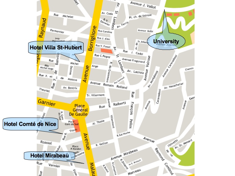
Accommodation
- Hôtel Comté De Nice **29, rue de DijonPhone: +33 4 93 88 94 56Web: http://www.hotelcomtedenice.com
- Villa Saint-Hubert **26, rue Michel-AngePhone: +33 4 93 84 66 51Web: http://www.villasainthubert.com
- Hôtel Mirabeau ***15 avenue MalaussénaTél. : 04 93 88 33 67Fax : 04 93 16 14 08
- Hôtel Le Floride **SG 43# / DBL 55 à 63#52, Bd de Cimiez-06000 NiceTél: 04 93 53 11 02Fax : 04 93 81 57 46
- Hôtel Villa Rose **SG 55# / DBL 58#43, Av de Bellevue-06000 NiceTél: 04 93 84 45 93http://www.cote.azur.fr/hotel_villa-rose-nice_521_lang_en.htm
Registration
Registration is free but mandatory : Workshop > Registration.
Directions
- Bus and tram website: public transport of Nice
- From railway station: see interactive map above.From railway station (“gare Thiers”) to campus (“Valrose”), you can go:
- by foot, approximately 30 minutes,
- by tramway (light rail).
In both cases, you have to exit the railway station, take “avenue Thiers” on the left, to the tramway stop.
If you choose the tramway option, direction “Las Planas”, and exit at stop “Valrose Université” (3rd stop). If you choose the foot option, turn to the left, and follow the tramway line (avenue Malaussenta, and then avenue Borriglione).
Once at “Valrose Université” stop, turn to the right, and then go straight to “avenue Joseph Vallot”. The campus is at the end of this street.
Map of the tramway line: http://tramway.nice.fr/La-ligne-1/Le-trace
- From airport to university, several options:
- There is a free (blue) shuttle from Terminal 2 to Terminal 1. From Terminal 1, take bus 23 (ticket = 1 euro), direction “Vallon des Fleurs”. Exit at “Joseph Vallot”, go back and turn left into “Joseph Vallot” street. The campus “Valrose”is at the end of this street. It takes 45 minutes – 1 hour.
- Faster: from terminal 1 or terminal 2, there is a fast shuttle, bus 98 (ticket = 4 euros), direction “Riquier”, and exit at “Cathédrale Vieille Ville” stop. With the same ticket, take the tramway direction “Las Planas” and exit at “Valrose University” stop. Turn right into avenue “Joseph Vallot”.
- Taxi: much faster, much more expensive (approx. 30 euros, 40 at night or week-end, even more if you look like a tourist…). Don’t forget to mention “Valrose” campus, as there are several university campus in the city.
- Hôtel Villa Saint-Hubert (NO LATE ARRIVAL! please contact us or the hotel in case of a late arrival, after 8pm):The hotel Saint-Hubert is located 26 rue Michel Ange, close to tramway stop “Borriglione”. To get there, see previous indications (airport/railway station to campus). Exit tramway at stop “Borriglione” (follow the tramway line and rue Michel-Ange is the next street, the hotel is half a block on the left); or exit bus 23 at stop “Eglise Jeanne d’Arc”. Go back and turn right into rue Michel Ange, the hotel is 2 blocks from the parking.
- Hôtel Comté de Nice:The hotel Comté de Nice is located 29 rue de Dijon, close to “Libération” tramway stop. To fet there, see previous indications. From railway station, by foot or tramway (1 stop), the hotel is near the intersection between “rue de Dijon” and “rue Clément Roassal” (walking street close to the tramway stop). From airport, tramway stop “Libération”, or bus 23, exit at stop “Libération”, turn to the next right and follow the tramway line, and turn right in “Rue Clément Roassal” (close to tramway stop), and then right in “rue de Dijon”.
- From hotels to Valrose campus:Get to avenue Malausséna (for Comté de Nice) or avenue Borriglione (for Villa Saint-Hubert), where the traway is. Follow the tramway line, direction “Las Planas” (to the left when you reach the tramway line). At station “Valrose Université” (1 stop from Borriglione and Villa Saint-Hubert, and 2 stops from Libération and Comté de Nice), turn right into avenue Joseph Vallot, the campus is at the end of the street.
Some more maps…:
- Map of campus and railway station
- Zoom on the campus
- Map of Valrose campus (conference location: maths department)